Corneal and Ocular Immunology Unit
Research Overview
Overwiew
Our research is aimed at understanding how corneal epithelial cells, immune cells and sensory nerves interact to achieve homeostasis and rapid recovery following injury, exposure to noxious stimuli and during normal ageing. Our laboratory is based in the Melbourne Brain Centre, where we have access to a suite of state-of-the-art instruments to enable in vivo clinical imaging of the mouse eye, along with ex vivo imaging of corneas, tissue culture and molecular biology techniques.
Corneal immune cells
The mouse and human cornea contains populations of resident immune cells such as macrophages and dendritic cells. These cells play an important role in generating innate inflammatory responses against microbial pathogens and sterile injurious stimuli, as well as interacting with the dense population of sensory nerves that supply the corneal epithelium.
Neuroimmune interactions
Recent studies suggest that corneal dendritic cells are closely associated with corneal sensory nerves. The nature of this relationship is thought to be spatial (i.e. the two entities are physically in contact), physiological (dendritic cells are thought to promote nerve homeostasis) and immunological (i.e. nerve stimulation affects dendritic cell activation). We have a range of active projects in the lab investigating the interplay of corneal sensory nerves and the effect that tissue injury and exposure to noxious stimuli has on the activation state of the resident immune cells.
Major techniques:
- In vivo clinical imaging of corneal inflammation using spectral domain optical coherence tomography
- Ex vivo confocal imaging of immunostained tissues, 3D reconstruction and image analysis
- Live cell imaging of corneal immune cells
- Gene and protein expression of chemokines, cytokines and neuropeptides
Other research interests:
- Membrane nanotubes in mammalian tissues and their responsiveness to stress signals.
- Distribution and phenotype of monocyte-derived cells in the mouse choroid plexus and meninges.
- Responsiveness of corneal nerve-associated macrophages to peripheral injurious stimuli.
- Retinal and intraocular inflammation as a result of corneal and anterior segment inflammatory events in the mouse eye.
Staff
Staff
Helen Jiao (Post - doctoral fellow)
Students
Manikkuwadura De Silva (PhD student)
Kirthana Senthil (Master of Biomedical Science student)
Mengliang Wu (PhD student)
Collaborators
- Dr Laura Downie, Department of Optometry and Vision Sciences, University of Melbourne
- Dr Lisa Hill, Institute of Inflammation and Ageing, University of Birmingham
- Prof Paul McMenamin, Department of Anatomy and Developmental Biology, Monash University
- Dr Samantha Dando, School of Biomedical Sciences and Institute of Health and Biomedical Innovation
Funding
This lab has been funded by the NH&MRC since 2013.
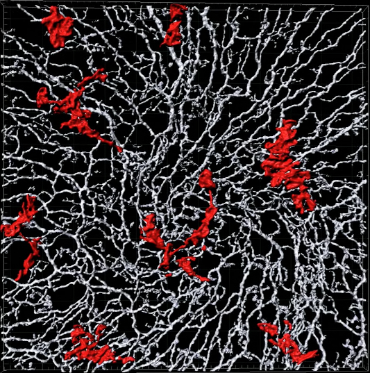 | Corneal macrophages (red) located beneath the epithelial |
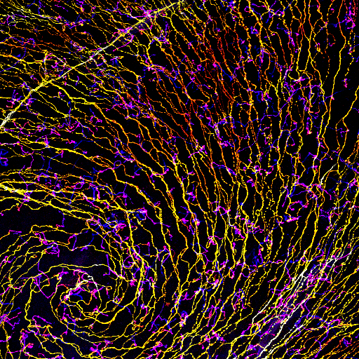 | A false coloured depth projection of the epithelial nerve plexus in the central cornea of a mouse. |
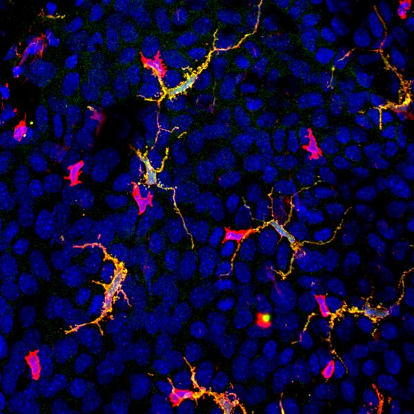 | (red) in the epithelium of the mouse cornea. |
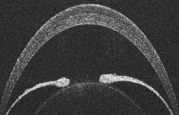 | A cross-sectional view of the mouse anterior segment imaged using spectral |