
Welcome to
Melbourne Eyecare Clinic
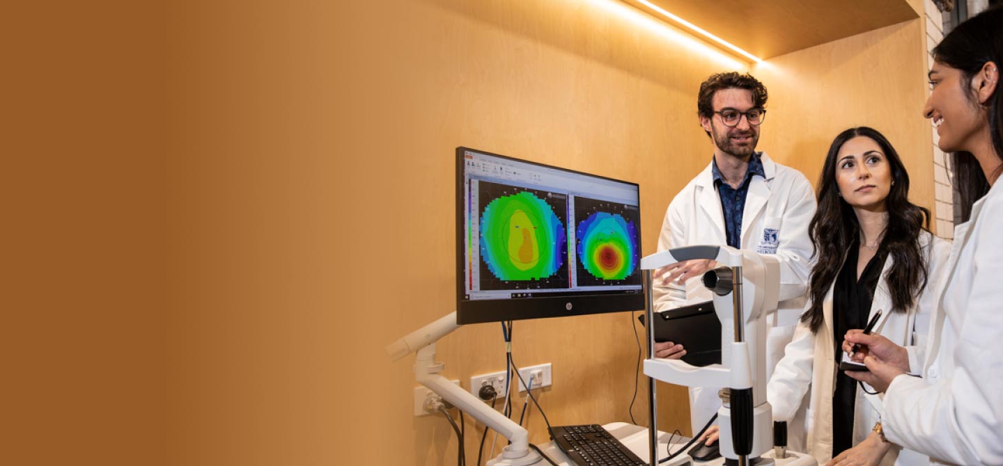
We are a University of Melbourne teaching clinic, centrally located in Carlton, providing a comprehensive suite of eyecare services to the public.
Our Services
Primary Eyecare | Cornea & Contact Lenses | Colour Vision | Children's & Binocular Vision | Dry Eye | Myopia | and much more.
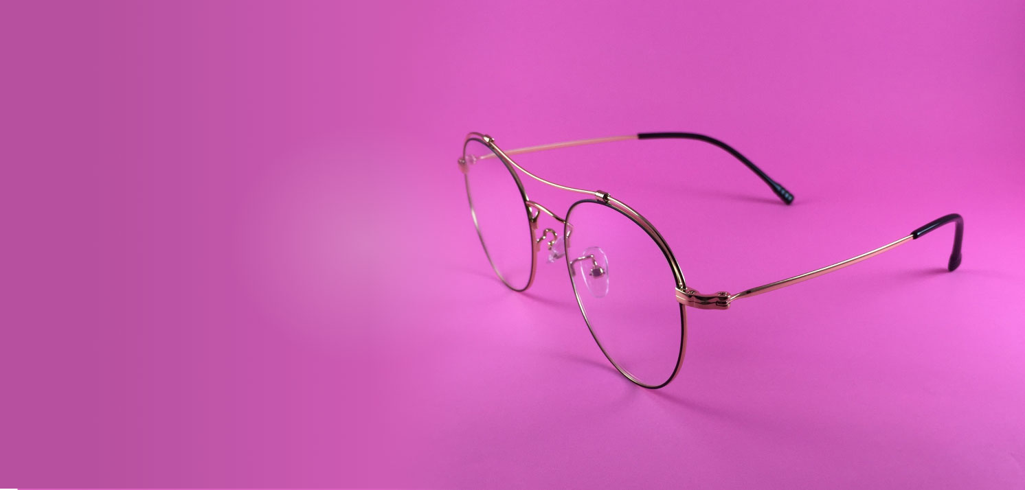
Eyewear
We have an extensive range of eyecare products like glasses, contact lenses and sunglasses.
Latest offers and quick links
Our Team

Contact Us
Contact Us
Melbourne Eyecare Clinic
200 Berkeley St,
Carlton 3053
Phone: 03 9035 6666
Email: uni-eyecare@unimelb.edu.au
Opening hours
Monday, Tuesday Thursday & Friday
8:30 am to 5.00 pm
Wednesday
8:30 am to 7.30 pm
Public holidays subject to change.